Originally published by ImageWorks Corporation.
Link to original article: https://www.imageworkscorporation.com/case-study-abscess-on-24/
By Dr. Paul Blaisdell, Kuna
I recently had an X-era Cone Beam from Imageworks installed in our office. Our staff was very excited to bring the new capability to our practice, and so far it has surpassed expectations by helping us provide a level of care for our patients that’s only possible with the information that 3D dentistry offers.
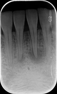
I wanted to share a unique case we’ve had with one of our patients. This patient presented for a routine periodic exam. We hadn’t taken anterior PAs prior to this appointment. However, upon seeing anterior PA’s, an apical radiolucency was noted on #24.

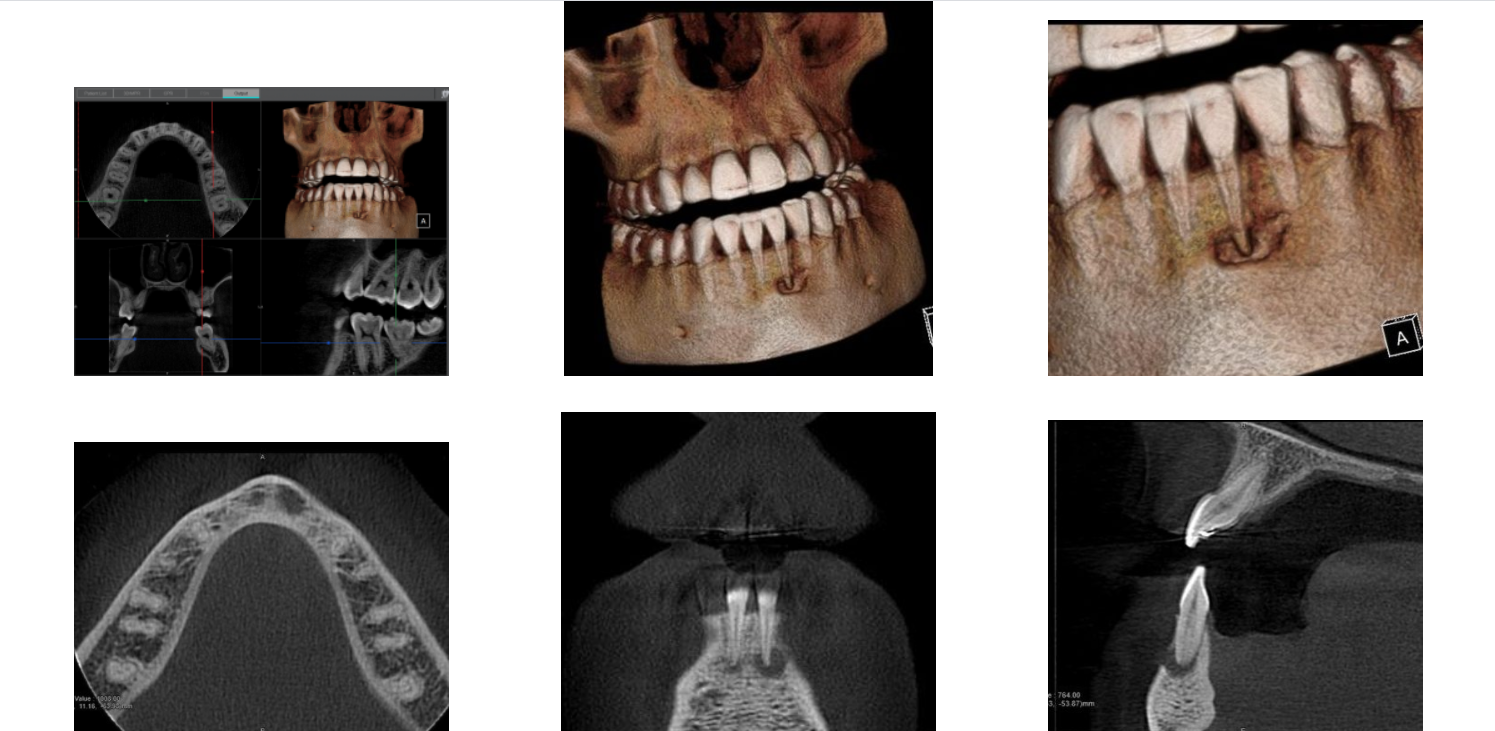
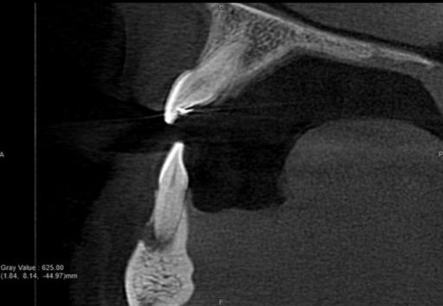
A CBCT was taken after a negative cold test indicated a necrotic tooth. When the CBCT was taken, we could clearly see that the abscess had completely perforated the labial bone at the apex of #24 and was close to doing so on #25.

As we investigated more deeply, we identified another critical piece of information that would affect our treatment plan: a second canal on #24.

Had I initiated treatment on this particular tooth and kept my access very conservative, there’s a chance I would have missed the other canal. Furthermore, had I started the treatment and then found the second canal after access into the chamber, I would have had to spend time determining the anatomy of both canals. For example, I would have had to determine if they had separate apicies.
The X-era from Imageworks helped me in a number of ways. First, it helped me feel very comfortable keeping this case in-house, which allowed me to keep the production. This was an immediate quantitative benefit that shows me the system will pay for itself over a short period of time.
Second, I was able to utilize the dramatic effect of such a high resolution volume to educate the patient in a clear and compelling way. I was able to stress the importance of taking care of this situation.

Finally, and most importantly, I’m confident that my proposed treatment plan is optimal for the patient in that it maximizes effectiveness while minimizing invasiveness.
This all took place just a couple for days after receiving our initial training. While I’m an enormous fan of the image quality and information that it provides, I think my staff’s favorite part is that the machine is so easy to use. Furthermore, the team at Imageworks have been fantastic to work with, as they’ve been able to help us with any questions we’ve had along the way.
Subscribe to Receive More Great Articles
